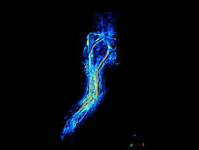
A pioneering technique developed by engineers at University College London (UCL,London,UK;www.ucl.ac.uk) that combines highly-detailed,real-time images of inside the body with a type of infrared light has been successfully used for the first time to differentiate between cancerous tumors and healthy tissue during surgery.
The engineers used the technique during preclinical testing in mice to successfully identify part of a tumor that had not been removed during surgery.
The technique may have significant implications for treating neuroblastoma in children,which is the most common form of solid cancer tumor,apart from brain tumors,found in children.